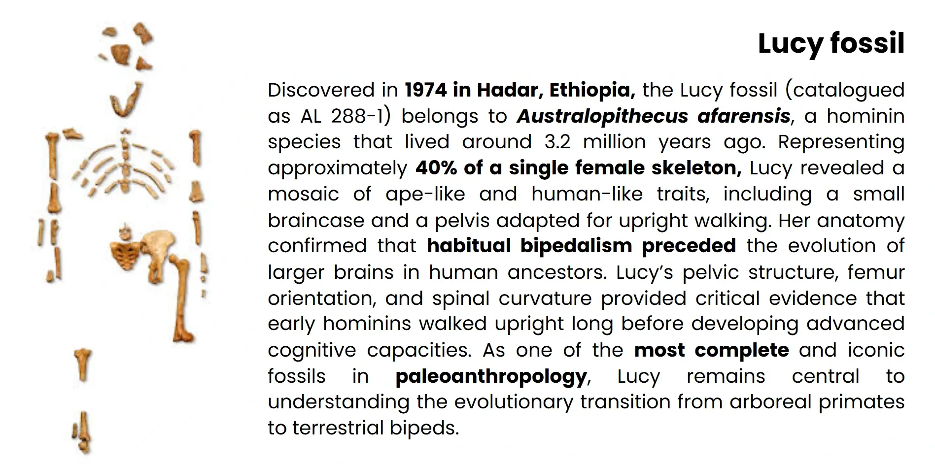
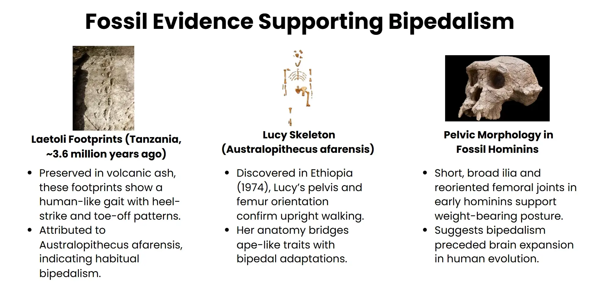
A new study shows that human bipedalism was achieved through two developmental changes-redirecting cartilage growth and delaying when bones are formed-both of which re-engineered the pelvis to support upright walking.
The advent of bipedalism is a landmark event in the history of hominids and a precursor of an anatomical, behavioural and cognitive superiority since the species has acquired a multitude of new faculties. Traditionally, the origin of upright gait has been blamed on environmental pressures and morphological adaptations as determined from fossils. Recent interdisciplinary studies, however, have shifted the focus to developmental biology with exciting evidence that the origins of human bipedalism include two different embryonic mechanisms: directed cartilage growth and delayed bone formation in the pelvis. These findings give conventional narratives to the contrary by emphasising internal developmental reprogramming rather than incremental anatomical change. The use of comparative embryological analysis and modern molecular techniques (e.g. spatial transcriptomics, single cell multi-omics) has revealed the role that these pelvic re-modelling played in enabling bipedal locomotion while remaining compatible with obstetric imperatives. This article describes the historical evolution of bipedalism, explains breakthrough research on the topic, and considers the larger evolutionary and medical implications of these developmental changes. Combining fossil data with phylogenetic and histological information, the study provides a new model for interpreting the acquisition of bipedalism in early hominins, as a result changing both evolutionary thought and biomedical research.
History of ManBipedalism
The origin of bipedalism represents a key transition in human evolution, bringing about profound changes in morphology, behaviour and ecological adaptation. Its causes are inseparably intertwined with environmental changes and selective pressures.
From Arboreal Locomotion to Terrestrial Adaptation
Early hominins evolved from trees-dwelling primates, whose morphology was adapted to life in the trees: long forelimbs, grasping feet and highly flexible spines were worker features that helped them arboreal-grasp and brachiate. Between about 7 and 10 million years ago, climate change in Africa caused dense forests to undergo a clumping into open woodlands and savannahs. The latter ecological shift minimised canopy cover and terrestrial exposure, forcing primates to evolve cutaneous Locomotor behaviours for movement on the ground.
Theories and fossil evidence
Among the various theories of bipedalismCharles Darwin has argued that it had something to do with the opportunity for the hands to be released from use for tools and for fighting, but archaeology suggests that tool production preceded the development of bipedal locomotion. Other hypotheses stress on aquatic wading, transportation of food or babies, and presentation. Archipelagos of study: Australopithecus afarensis's pelvis, particularly the "Lucy" specimen, and the Laetoli footprints provided us with anatomical evidence of habitual upright walking, such as flared ilia and reoriented femoral joints.
Evolutionary Significance
Bipedalism was more than just a biomechanical change; it itself was a force for evolutionary change. It has changed the structure of the axial skeleton, lower limb and hence resulted in an impact on the Childbirth and social behaviour. Later hominin characteristics, including cognitive ability expansion,manual agility and cultural innovation were made possible by the upward ambulation. As Capellini has suggested, clarifying the developmental processes that underpin bipedalism sets forth a re-assessment of long standing notions of human origins.
The Study's Breakthrough
Publishing in Nature, an interdisciplinary study has clarified our knowledge of the evolution of bipedalism (walking), identifying two developmental innovations in pelvic anatomy that allowed for erect gait among archaic humans.
Unusual growth of cartilage in the ilium
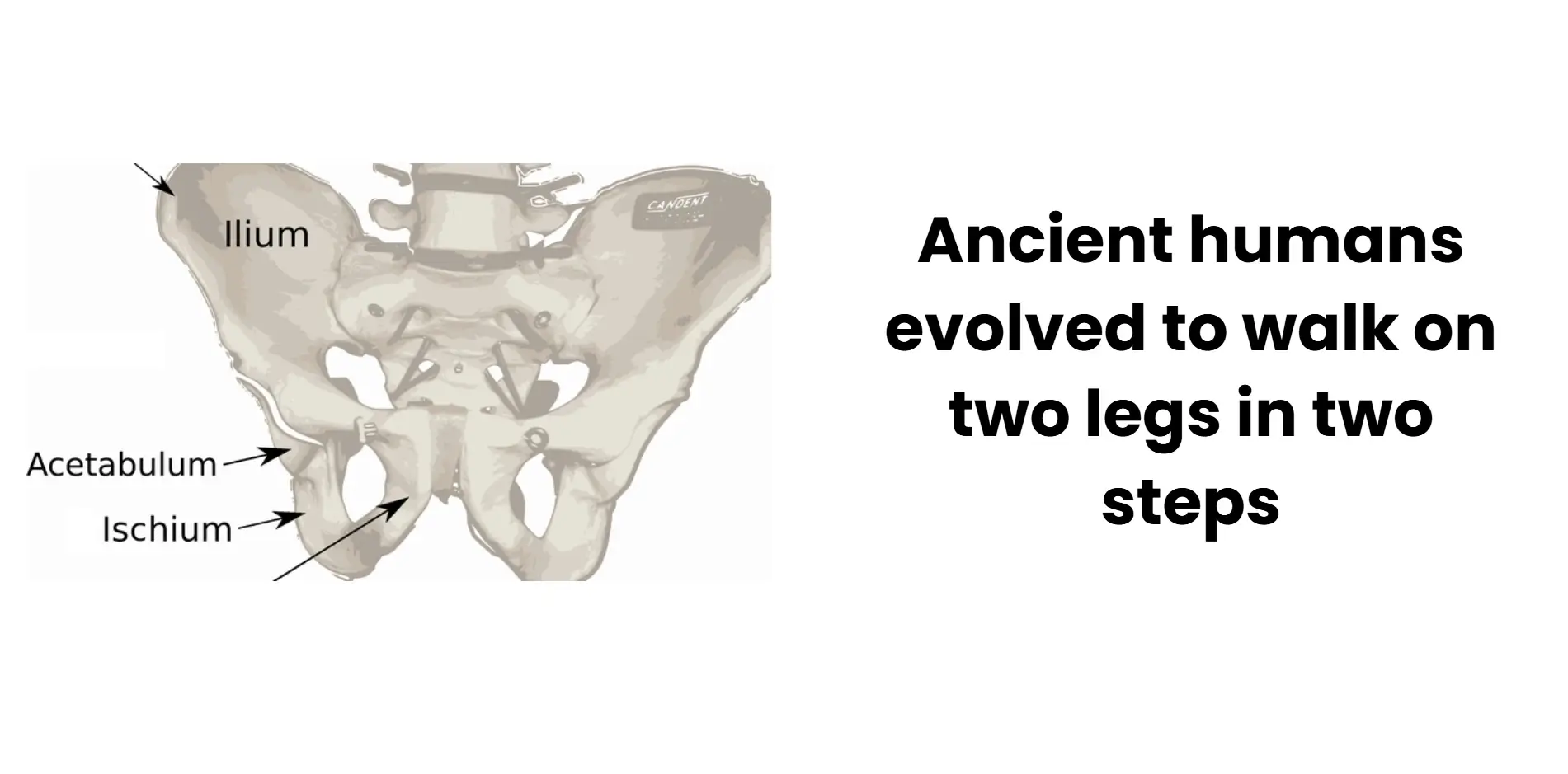
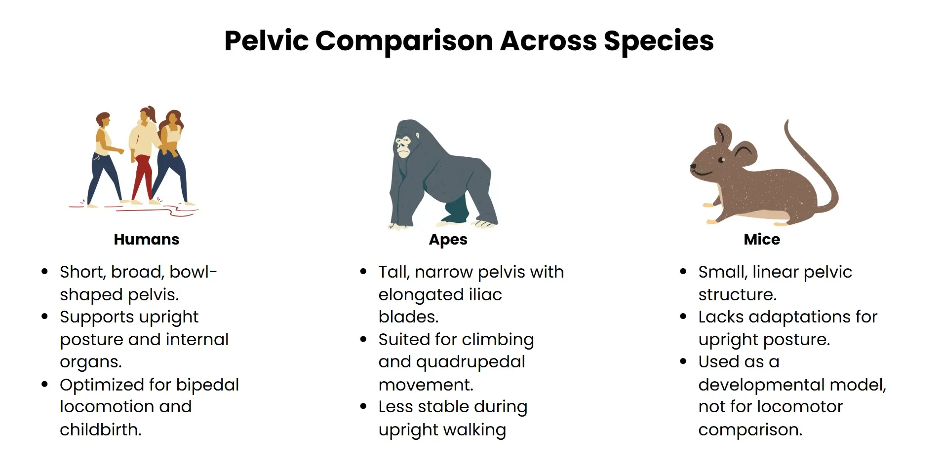
The first major finding is in iliac cartilage. In contrast with apes and rodents, which express a mainly vertical elongation of cartilage, human embryos display a lateral extension of the iliac growth plate. This change results in a shorter, wider bowl-shaped pelvis that is structurally adapted for vertical movements-optimised for balance and load shifting during bipedal locomotion. To elucidate this process, the researchers used the techniques of histology and micro-CT imaging of crucial developmental phases in embryos and compared human samples with those of chimpanzees, gibbons, and mice.
Delayed ossification and Pelvic flexibility
The second arrives at the subject of timing and space involvement of bone formation. In humans, the ilium ossifies inward rather than outward, starting later and exogenously at the posterior margin. This delay allows the pelvis to increase lateral dimensions prior to calcification, an adaptation that is useful not only for locomotion, but for deliverance by big-brained infants. Using single-cell multi-omics and spatial transcriptomics profiling, the researchers found these changes are controlled by a complex regulatory network of genetic switches instead of a single "bipedalism gene."
Implications for Evolution and medicine
These results indicate that bipedalism was achieved by a two-step reprogramming of the development of the pelvis by reassignment of cartilage development and delayed ossification. This mechanistic sack shows us why fossil hominins such as Australopithecus already had short broad pelvis at millions of years ago. In addition, understanding these developmental pathways may shed light on the etiologies of pelvic malformations and skeletal disorders in modern humans.
Step-1: Redirected Cartilage Growth
Recent work on development has highlighted an anatomical niche change in early hominin evolution: the reversal of growth direction of iliac cartilage that essentially reorganised the pelvis to support bipedalism.
Horizontal Growth Plate Iliac Expansion
In most primates the iliac cartilage (the superior part of the pelvis) extends upward in length, giving rise to a tall and narrow morphology adapted to quadrupedal or arboreal locomotion. In the human embryo, on the other hand, it is the horizontal expansion of this growth plate which characterises the short and broad ilium. In the study led by researchers at Harvard and published in the journal Nature, the team used histological mapping and micro-CT imaging to compare pelvic development during embryology in humans, chimpanzees, gibbons, and mice.
Developmental and Evolutionary Innovation
The redirection of cartilage is a developmental originality and not a morphological sequence. Unlike other skeletal features, in non-human primates there are no evolutionary intermediates in the ilium. Spatial transcriptomics and single-cell multi-omics analysis unravelled a network of transcriptional switches active during embryogenesis that likely regulates horizontal growth pattern. These molecular data suggest that bipedalism evolved not only by means of extrinsic environmental forces but through endogenous developmental reprogramming.
Functional Relevance to Bipedalism
The wider pelvis generated from shift to preload cartilage growth offers essential support for upright posture, internal organ stability and Locomotor efficiency. It also makes it possible to keep the gluteal muscles in place needed for balance and stride control. This skeletal change paved the way for later morphological features including adjustments in axial curvature of the spine and the positioning of lower limb segments, further support was provided for pelvic construction centrality to early human bipedalism.
Step-2: Delayed Bone Formation
Among the evolutionary novelties associated with human bipedalism is late skeletal maturity of the pelvic skeleton, a developmental change that maintained pelvic stability and facilitated upright locomotion and effective childbirth.
Posterior mode of ossification
Unlike in other primate species where formation of the pelvic bone also starts early and progresses centrally through cartilage, ossification of the pelvic bone has a delayed onset of embryonic development in humans. This temporal lag allows a horizontal widening of the cartilaginous precursor and then mineralization occurs, maintaining the short broad morphology necessary for bipedal stability. Geometric morphometric techniques based on micro-CT scans and histological description confirmed this ossification pattern through the chronological phases, confirming a different sequence than in chimpanzees and gibbons.
Epigenetics, Regulation and Developmental Re-Programming
This temporal pattern of bone formation is controlled by the complex construction of a network of genetic regulators instead of a single gene. Comprehensive single cell and spatial transcriptomics and single cell multi-omics identified hundreds of regulatorymodules coordinating the delayed ossification time course. Critical transcription factors SOX9 and parathyroid hormone-related protein (PTHrP) were identified as the modulators of cartilage cell differentiation and bone maturation. These molecular mechanisms highlight how evolutionary novelty may develop by fine-tuned changes and the modulation of developmental time and space of where a gene is expressed.
Functional and Evolutionary Consequences
Slowly evolving osteogenesis not only allowed human bipedalism but also solved an obstetric challenge-extensive craniofacial accommodation to carry a large brain infant. Retardation of ossification allowed the pelvis to remain flexible during gestation and childbirth. This dual selection advantage i.e. locomotor efficiency combined with an advantage toward high reproductive success, informs a central role for delayed ossification in hominin evolution. It also yields information on congenital pelvic disorders and skeletal deformities in modern man.
Evolutionary and Medical Significance
For example, the developmental progression of pelvic cartilage proliferation and ossification timing have not only shaped the locomotory patterns of humans, but also produced profound consequences for evolutionary fitness and modern medical dogmas.
Re-shaping Evolutionary Trajectories
Hominin body schema was redrawn with the development of an adaptive bipedalism due to modified cartilage extracellular matrix production with rutting temporality of ossification. The coronal curving morphology of an anthropomorphic pelvis allowed bipedalism, freeing the upper limbs to manipulate hands and other tools, interact with each other, and process the environment. Such morpho-functional adjustments further facilitated long-distance dispersal and thermoregulatory effectiveness which enabled the success of early humans in a breadth of climatic and topographic regimes. Importantly the increased width pelvic supported the birth of large brained infant and linking locomotor evolution and cognitive expansion. The result for this was a dual trait experience (mobility and reproduction) that represented a turning point in the success of hominins in the terrestrial biomes.
Developmental Insights into Skeletal Disorders
Furthermore, understandings of the mechanisms that underpin pelvic remodelling have brilliantly illuminated disease mechanisms of the congenital and degenerative pulmonary compartments. Although delayed ossification was seen as providing selective advantage, there is a possibility that delayed ossification also makes affected individuals more susceptible to hip dysplasia, pelvic asymmetry, and other associated pathology. By whole-genome spatial transcriptomics profiling and single cell multiomics analyses we have identified regulatory network co-regulation of bone accrual underpinning future therapeutic strategies in skeletal malformations and growth disorders. Moreover, the findings obtained should help in refining orthopaedic protocols and in adjusting prenatal diagnostic algorithms by identifying critical (time) stenosis periods in pelvico-axial morphogenesis.
Detention of Evolution and Biomedicine
This question reflects the translational value in the application of evolutionary biology to clinical practise. By resolving the origin of human bipedalism to the discrete embryological reconfiguration, the investigators are able to better specify the anatomical susceptibilities inherent to morphology. The pelvis, once the evolutionary innovation, has, now turn in afocal point for the development of solutions to modern health problems from childbirth complications to advancing degenerative joint disease.
Conclusion
The phylogenetic origin of human bipedalism represents a profound anatomical and developmental transformation, and reflects not solely extrinsic ecological pressures, but rather the intrinsic effects of a change in the mechanics of embryonic growth. The recent articulation of two of the principal mechanisms like redirection of cartilage proliferation and delayed ossification provides a strong developmental paradigm for tackling the evolutionary scenario of human pelvic morphogenesis for upright locomotion. These results connect gaps between fossil morphology, genetic regulatorycircuitry and functional anatomy, which thereby re-contextualize long-standing narratives within paleo-anthropology. Moreover, the implications are not confined to the borders of the evolutionary biology and add worthily to contemporary biomedical efforts in coping with skeletal disorders and developmental aberrations. Describing the molecular and mechanical substrates underlying pelvico-axial remodelling, this work emphasises the potential of interdisciplinary thinking in understanding the ontogeny of complex traits. Ultimately, this work furthers our understanding of the multiple pathways of human evolution and helps to affirm the dynamic interrelation between form, function, and the chronology of evolutiondevelopment in that determine the human phenotype.